


When Do Babies Start to Recognize and Remember Their Mothers After Delivery?
 01 September,2025
Read More
01 September,2025
Read More
Enquire now in case of any assistance needed

Slit Lamp Examination Treatment Cost in India is between USD 30 - USD 100

Hospital Days: 1

Procedure Duration: 10 Min - 20 Min
The slit lamp examination, a cornerstone of eye care, provides detailed insights into ocular health. Utilizing a binocular microscope with an adjustable light source allows clinicians to examine the eye's anterior and posterior segments. By illuminating the eye with a narrow beam of light, the slit lamp enables visualization of structures like the cornea, iris, lens, and retina with exceptional clarity. This examination aids in diagnosing conditions such as cataracts, glaucoma, and macular degeneration, guiding treatment decisions effectively. Its versatility and precision make it indispensable in routine eye examinations and specialized procedures, ensuring comprehensive and accurate ocular assessments.
A slit lamp examination is a vital component of comprehensive eye care, offering detailed insights into the health of various ocular structures. Here are reasons why a slit lamp examination is necessary in the generic form
In the field of ophthalmology, various types of slit lamp examinations are utilized to assess different aspects of ocular health. Here are some common types in a generic form:
Several factors influence the cost of a slit lamp examination, reflecting the complexity and range of services provided during the examination:
The selection of patients for a slit lamp examination is based on various factors, and eye care professionals consider specific signs and indications to determine the necessity of this examination. Here are common considerations:
Several diagnostic tests and evaluations are performed to determine the need for a slit lamp examination. These assessments help eye care professionals identify specific signs and indications, guiding them in deciding whether a more detailed examination is warranted:
1. Early Detection of Eye Conditions: Slit lamp examinations enable early detection of various eye conditions, including corneal abnormalities, cataracts, conjunctivitis, and anterior uveitis. Timely identification allows for prompt intervention and effective management.
2. Contact Lens Assessment: For contact lens wearers, slit lamp examinations play a vital role in assessing lens fit, identifying potential complications, and ensuring overall ocular health related to contact lens use.
3. Accurate Prescription for Eyeglasses or Contacts: The examination helps eye care professionals determine the accurate prescription for eyeglasses or contact lenses. This ensures optimal visual acuity and reduces eyestrain associated with refractive errors.
4. Monitoring Ocular Health: Slit lamp examinations are instrumental in monitoring the overall health of the eyes, providing insights into changes in the anterior segment structures and guiding long-term eye care.
1. Discomfort during Examination: Some individuals may experience mild discomfort during a slit lamp examination, particularly if eye drops are used for pupil dilation or fluorescein staining. However, any discomfort is generally short-lived.
2. Light Sensitivity: Pupil dilation during the examination may result in temporary light sensitivity, requiring individuals to wear sunglasses immediately afterwards. This effect diminishes as the pupils return to their normal size.
3. Rare Complications: While extremely rare, there is a minimal risk of adverse reactions to eye drops or unforeseen complications during the examination. However, these instances are uncommon and usually minor.
4. False Positives or Negatives: Like any diagnostic test, slit lamp examinations may have limitations, leading to false positives or negatives. Clinical judgment and follow-up assessments help mitigate potential discrepancies.
The benefits of slit lamp examinations, including early detection and accurate prescriptions, outweigh potential risks, which are generally minimal and transient. Regular slit lamp assessments contribute to maintaining optimal eye health, enabling timely interventions and enhancing overall well-being.
Recovery and rehabilitation after a slit lamp examination are typically straightforward, as the procedure is non-invasive and minimally discomforting. Here are key aspects of the recovery process:
1. Immediate Post-Examination Period: Individuals may experience mild discomfort during the slit lamp examination, particularly if eye drops were used for pupil dilation or fluorescein staining. However, any discomfort is generally short-lived.
2. Resolution of Temporary Effects: Light sensitivity due to pupil dilation or residual fluorescein staining usually subsides within a few hours. Vision returns to normal as these temporary effects wear off.
3. Resumption of Normal Activities: In most cases, individuals can resume their normal activities immediately after the slit lamp examination. There are usually no restrictions on daily routines or work-related tasks.
4. Adaptation to New Prescriptions: If the examination changes the prescription for eyeglasses or contact lenses, individuals may go through an adjustment period. Adapting to new lenses is natural and varies among individuals.
5. No Physical Recovery Required: Given that the slit lamp examination is non-invasive, there is no physical recovery needed. Patients do not experience postoperative pain or restrictions on movements.
6. Follow-Up Recommendations: Depending on the examination findings, the eye care professional may recommend follow-up appointments, additional tests, or specific interventions if necessary.
7. Communication with Eye Care Professionals: If individuals experience persistent discomfort or changes in vision after the slit lamp examination, it is important to communicate these concerns with the eye care professional promptly. This ensures timely intervention or clarification of any issues.
Recovery and rehabilitation after a slit lamp examination involve a brief period of adaptation to any temporary effects, with individuals usually returning to their normal activities shortly after the procedure. Open communication with the eye care professional ensures a smooth post-examination experience and addresses concerns.
After a slit lamp examination, patients can generally expect minimal discomfort or side effects. The procedure itself is painless and non-invasive, involving the use of a microscope-like device called a slit lamp to examine the eyes' anterior and posterior segments. Here's what patients may experience after the examination:
1. Performing a Slit Lamp Examination: A slit lamp examination is a precise and comprehensive assessment of the anterior segment structures of the eye. Here's an overview of how the examination is typically performed:
2. Patient Preparation: The patient is seated comfortably behind the slit lamp biomicroscope, and their forehead and chin are rested on supports to maintain stability during the examination.
3. Adjustment of the Slit Lamp: The eye care professional adjusts the height and position of the slit lamp to align it with the patient's eyes. Proper alignment ensures a clear and detailed view of the eye structures.
4. Illumination and Magnification: The slit lamp combines a bright light source with a microscope that provides high magnification. This allows the eye care professional to thoroughly examine the cornea, iris, lens, and anterior chamber.
5. External Eye Examination: The examination begins with assessing the external eye structures, including the eyelids, conjunctiva, and lacrimal glands. Any signs of inflammation, infection, or abnormalities are noted.
6. Corneal Examination: The slit lamp is focused on the cornea to assess its clarity, curvature, and the presence of any abnormalities, such as scratches, scars, or foreign bodies.
7. Iris and Pupil Assessment: The iris and pupil are examined for size, shape, and irregularities. The slit lamp helps detect conditions like iritis or anisocoria (unequal pupil size).
8. Lens Examination: The lens is carefully examined for clarity and any signs of cataracts. Changes in lens opacity or position can be identified during this part of the examination.
9. Anterior Chamber Evaluation: The anterior chamber, the space between the cornea and iris, is assessed for depth and signs of inflammation or abnormalities.
10. Tonometry - Eye Pressure Measurement: In some cases, tonometry may be performed during the slit lamp examination to measure intraocular pressure, a key factor in assessing the risk of glaucoma.

Consultant
Cardiothoracic and Vascular Surgeons
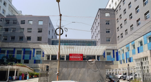
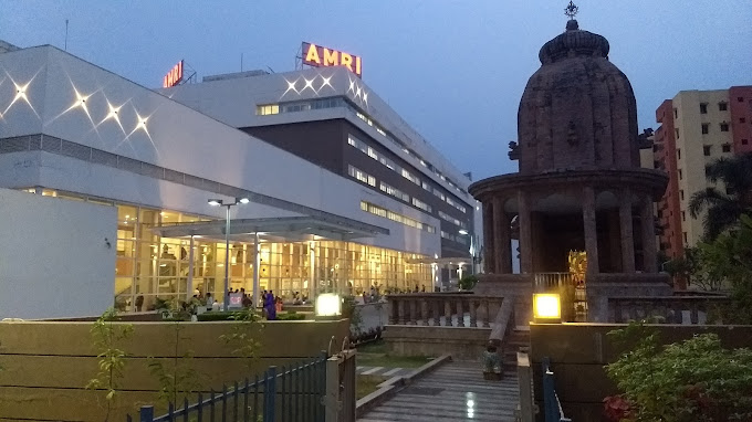
Apollo Gleneagles Medical Centre, Kolkata
View Doctor Profile Book an Appointment
Senior Consultant
Neurologists
Calcutta Medical Research Institute | CK Birla Medical Centres, Kolkata
View Doctor Profile Book an Appointment
Consultant
Ophthalmologist
Calcutta Medical Research Institute | CK Birla Medical Centres, Kolkata
View Doctor Profile Book an Appointment
Consultant
Ophthalmologist
Calcutta Medical Research Institute | CK Birla Medical Centres, Kolkata
View Doctor Profile Book an Appointment
Consultant
Ophthalmologist
Calcutta Medical Research Institute | CK Birla Medical Centres, Kolkata
View Doctor Profile Book an Appointment


Senior Consultant
Medical Oncologists
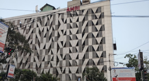
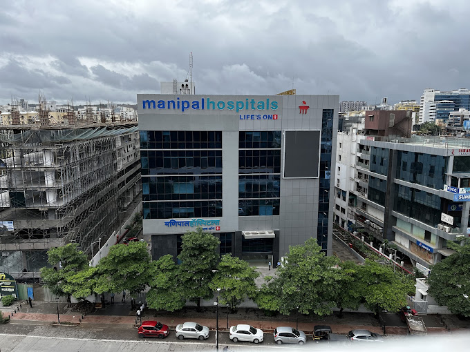
Manipal Medical Centre Formerly AMRI Medical Centre, Dhakuria, Kolkata
View Doctor Profile Book an Appointment
Doctor of Pharmacy
Dr. Deepanshu Siwach is a skilled clinical pharmacist with a Doctor of Pharmacy degree. He has 4+ years of experience and has worked with thousands of patients. He has been associated with some of the top hospitals, such as Artemis Gurgaon and Teerthanker
Dr. Deepanshu Siwach is a skilled clinical pharmacist with a Doctor of Pharmacy degree. He has 4+ years of experience and has worked with thousands of patients. He has been associated with some of the top hospitals, such as Artemis Gurgaon and Teerthanker...
The Art of Effective Communication
 01 September,2025
Read More
01 September,2025
Read More
 01 September,2025
Read More
01 September,2025
Read More
 01 September,2025
Read More
01 September,2025
Read More
 01 September,2025
Read More
01 September,2025
Read More
 01 September,2025
Read More
01 September,2025
Read More
 31 August,2025
Read More
31 August,2025
Read More
 31 August,2025
Read More
31 August,2025
Read More
 31 August,2025
Read More
31 August,2025
Read More
 31 August,2025
Read More
31 August,2025
Read More
 31 August,2025
Read More
31 August,2025
Read More